EMDR Research News May 2014
Among the five EMDR therapy articles, there are two case reports. These examine EMDR therapy for mild dementia and for complex regional pain syndrome. There is a research report from the Netherlands showing that the eye movements used in EMDR therapy produce enduring changes in memory but only when there is sufficient duration of the eye movements. There is a Clinical Q & A article on a new metaphor for helping clients understand their issues and recognize their gains in the EMDR approach to therapy. Finally, there is a clinical overview of how to apply EMDR therapy to cases of borderline personality disorder from my Spanish co-authors, Dolores Mosquera and Anabel Gonzalez and myself.
In addition you will find a summary of a French article on magneto-encephalographic (MEG) brain recordings during traumatic memory recall in women with post-traumatic stress disorder which is dedicated to the memory of our colleague David Servan-Schreiber who died in 2011. Finally, there is full text link to an in-depth article on a theoretical model of the neurobiology of the hyperarousal subtype of PTSD.
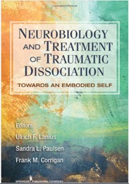
In the books section you will find the just released book on EMDR Therapy from Ulrich F. Lanius PhD, Sandra L. Paulsen PhD, and Frank M. Corrigan MD - Neurobiology and Treatment of Traumatic Dissociation: Towards an Embodied Self and a preview of the forthcoming EMDR Toolbox: Theory and Treatment of Complex PTSD and Dissociation from Jim Knipe, Ph.D.
With each reference below, you will find the citation, abstract and author contact information (when available). Prior quarterly summaries of journal articles can be found on the EMDRIA website and a comprehensive listing of all EMDR-related research is available at the Francine Shapiro Library. EMDRIA members can access recent Journal of EMDR Practice and Research articles in the member’s area on the EMDRIA website. JEMDR issues older than 12 months are available open access on IngentaConnect.
---
Recently released and forthcoming books on EMDR

.
---
Journal Articles
---
Amano, T., & Toichi, M. (2014). Effectiveness of the on-the-spot-EMDR method for the treatment of behavioral symptoms in patients with severe dementia. Journal of EMDR Practice and Research, 8(2), 50-65. doi:10.1891/1933-3196.8.2.50
Tamaki Amano, Yoshida Konoe-cho, Sakyo-ku, Kyoto 606-8501, Japan. E-mail: yiu60432@nifty.com
Abstract
Although the main symptoms of dementia consist of neuropsychological impairment, particularly long-term memory, dementia often involves severe behavioral and psychological symptoms of dementia (BPSD). There are quite a few patients whose BPSD are untreatable with medication. Such BPSD often have some characteristics similar to traumatic symptoms and appear related to the recollection of disturbing past traumatic events. Because the standard protocol of eye movement desensitization and reprocessing (EMDR) is not directly applicable to patients with dementia, we developed a modified protocol, the on-the-spot-EMDR method. This study describes the protocol and evaluates its application to three patients with moderate to severe dementia. Clear therapeutic effects were evident, and all three individuals showed pronounced improvement in BPSD, with results maintained at 6-month follow-up. The relevance of these findings is discussed and suggestions made for future research.
---
Cottraux, J., Lecaignard, F., Yao, S. N., De Mey-Guillard, C., Haour, F., Delpuech, C., & Servan-Schreiber, D. (2014). [Magneto-encephalographic (MEG) brain recordings during traumatic memory recall in women with post-traumatic stress disorder: A pilot study.]. L'Encephale. doi:10.1016/j.encep.2014.03.002
J. Cottraux, Unité de traitement de l’anxiété, service de psychologie médicale, hôpital neurologique, 59, boulevard Pinel, 69677 Bron, France. cottraux@univ-lyon1.fr
This work is dedicated to the memory of our friend David Servan-Schreiber (1961-2011).
Summary
Aim of the study: The experiment studied the effects of a short duration exposure to traumatic memories using magneto-encephalography (MEG).
Patients: Nine right-handed DSM-4 PTSD patients were recruited from a unit for anxiety disorders and an organisation supporting victims of violence. In order to have a homogeneous sample, we included only women who suffered from civilian PTSD. Exclusion criteria were co-morbid major medical illness, metallic dental prostheses that would interfere in the magnetic measurement, and current drug treatment. All participants were free from neurological disease and had normal hearing. They signed a written informed consent form. An ethics committee accepted the study.
Method: A tape-recorded voice administered a script-driven imagery. The patients had to imagine, successively, a neutral image, a traumatic memory and rest, while MEG measured brain activities across delta, theta, alpha and beta bands. Each condition lasted three minutes. Heart rate (HR), anxiety and the vividness of mental images were recorded at the end of each phase. MEG power analysis was carried out with Statistical Parametric Mapping (SPM) 8. The signals were averaged for each of the three conditions of three minutes duration. The dependent variable was a subtracted value: (trauma – rest) − (neutral – rest). The significance threshold was set at P < 0.01.
Results: Anxiety and HR significantly increased during the trauma condition and returned to the neutral level during rest. The vividness of the mental imagery remained stable across the three conditions. The left-brain demonstrated a statistically significant power decrease in the secondary visual cortex (BA 18-19) in the delta band, the insula (BA13) in the beta band, the insula (BA13), premotor cortex (BA 6), Broca area (BA 44), and BA 43, in the alpha band.
Discussion: The symptom provocation protocol was successful in eliciting subjective anxiety and HR response in relation to traumatic memories. Our MEG results are in keeping with previous neuro-imagery studies showing decreased activities in the insula and Broca area during PTSD symptom provocation. However, we did not replicate the activation in the amygdala and the cingulate and prefrontal cortex found in some studies. Moreover, the within-group design, the small sample, and the inclusion of only female patients with milder dissociative symptoms limit our conclusions. The MEG protocol we used may also explain some partial discrepancies with previous MEG studies. However, our aim was to provoke a specific autobiographic recall of a traumatic event unfolding several sequential mental images along three minutes as in exposure therapy for PTSD.
Conclusion: Despite its limitations, this pilot study is the first to provide MEG data during trauma recall. It suggests that recalling a specific traumatic event along three minutes results in hypo-activations of the brain regions regulating language and emotions. This paves the way to recording whole sessions of specific therapies for PTSD, with MEG using the millisecond resolution. MEG might be of interest to study the suppression of traumatic memories and their activation and habituation through prolonged graduated exposure in imagination across several sessions. MEG could also be used to study the effects of medication on PTSD symptoms. A controlled replication in a larger sample including male and female patients with various traumatic experiences is needed.
---
Hughes, M. (2014). EMDR as a therapeutic treatment for complex regional pain syndrome: A case report. Journal of EMDR Practice and Research, 8(2), 66-73. doi:10.1891/1933-3196.8.2.66
Megan Hughes, MA, Registered Clinical Counsellor, BCACC, #1834, 301-1055 W. Broadway, Vancouver, BC, V6H 1E2 Canada. E-mail: meganhughes@vancpm.com
Abstract
Complex regional pain syndrome (CRPS) is characterized by ongoing pain, swelling, and stiffness following an acute injury. CRPS is difficult to diagnose, significantly impacts functioning, and is frequently incurable. Current treatments are pharmacotherapy, surgery, and physiotherapy. This case report describes the use of eye movement desensitization and reprocessing (EMDR) in the psychotherapeutic treatment of a woman diagnosed with CRPS in 2009 as a result of injuries sustained during an assault in 2004. This article reports on EMDR treatment provided 1–2 years after her diagnosis. At initial assessment, the client was debilitated and suicidal, unable to work or care for her children, and dependent on her family for financial support because of CRPS. Two phases of 7 EMDR sessions were provided; the first focused on past traumatic experiences; the second addressed her pain with Grant's (2009) EMDR chronic pain protocol. At the end of treatment, the client reported decreased pain, decreased substance dependence, improved mood and outlook, and was able to resume part-time work. Results were maintained at 8-month follow-up and suggest that EMDR was helpful for this client in reducing the symptoms associated with CRPS.
---
Jarecki, K. (2014). The seed-to-weed technique: Graphically illustrating symptom etiology, treatment, and resolution. Journal of EMDR Practice and Research, 8(2), 90-100. doi:10.1891/1933-3196.8.2.90
Kriss Jarecki, 552 Linden Ave., East Aurora, NY 14052. E-mail: harmonyhearth@aol.com
Abstract
This Clinical Q&A article explains the seed-to-weed technique. This strategy offers an opportunity to help a client understand the problem and treatment approach and the ability to see progress in treatment. These crucial areas are important to treatment engagement and success, whether working with children or adults. An illustrated garden metaphor is used to guide a client to look at his or her life experiences and gain an understanding of how events have contributed to the problems and concerns that bring them into therapy. The seed-to-weed technique provides a graphic means of presenting trauma, a treatment plan, introducing eye movement desensitization and reprocessing (EMDR), and tracking treatment progress. This article introduces and demonstrates the seed-to-weed technique.
---
Leer, A., Engelhard, I. M., & van den Hout, M. A. (2014). How eye movements in EMDR work: Changes in memory vividness and emotionality. Journal of Behavior Therapy and Experimental Psychiatry, 45(3), 396-401. doi:10.1016/j.jbtep.2014.04.004
Arne Leer, Department of Clinical & Health Psychology, Utrecht University, P.O. Box 80140, 3508 TC Utrecht, The Netherlands. E-mail: A.Leer@uu.nl
Abstract
BACKGROUND AND OBJECTIVES: Eye movements (EM) during recall of an aversive memory is a treatment element unique to Eye Movement Desensitization and Reprocessing (EMDR). Experimental studies have shown that EM reduce memory vividness and/or emotionality shortly after the intervention. However, it is unclear whether the immediate effects of the intervention reflect actual changes in memory. The aim of this study was to test whether immediate reductions in memory vividness and emotionality persist at a 24 h follow up and whether the magnitude of these effects is related to the duration of the intervention.
METHODS: Seventy-three undergraduates recalled two negative autobiographical memories, one with EM ("recall with EM") and one without ("recall only"). Half of participants recalled each memory for four periods of 24 s, the other half for eight periods of 24 s. Memory vividness/emotionality were self-rated at a pre-test, an immediate post-test, and a 24 h follow-up test.
RESULTS: In both duration groups, recall with EM, but not recall only, caused an immediate decrease in memory vividness. There were no immediate reductions in memory emotionality. Furthermore, only the 'eight periods' group showed that recall with EM, but not recall only, caused a decrease in both memory emotionality and memory vividness from the pre-test to the follow-up.
LIMITATIONS: Only self-report measures were used.
CONCLUSIONS: The findings suggest that recall with EM causes 24-h changes in memory vividness/emotionality, which may explain part of the EMDR treatment effect, and these effects are related to intervention duration.
---
Mosquera, D., Leeds, A. M., & Gonzalez, A. (2014). Application of EMDR therapy for borderline personality disorder. Journal of EMDR Practice and Research, 8(2), 74-89. doi:10.1891/1933-3196.8.2.1
Dolores Mosquera, INTRA-TP, Instituto para el Es-tudio del Trauma y los Trastornos de la Personalidad, General Sanjurjo 111, 5° 15006, A Coruña, Spain. E-mail: doloresmosquera@gmail.com
Abstract
There is a growing interest in the use of eye movement desensitization and reprocessing (EMDR) therapy beyond posttraumatic stress disorder (PTSD) where its application is well established. With strong scholarly consensus that early traumatic and adverse life experiences contribute to the development of borderline personality disorder (BPD), EMDR would appear to offer much to the treatment of persons with BPD. However, given the specific characteristics of these clients, the application of EMDR therapy to their treatment can be challenging and necessitates several minor adaptations of the standard EMDR procedures for PTSD. This article provides an orientation to principles and strategies for safely and effectively preparing clients with BPD for EMDR therapy and for accessing and reprocessing the traumatic origins of BPD. Clinical examples are provided throughout.
---
Weston, C. S. (2014). Posttraumatic stress disorder: A theoretical model of the hyperarousal subtype. Frontiers in Psychiatry / Frontiers Research Foundation, 5, 37. doi:10.3389/fpsyt.2014.00037
Full text available in PubMed Central or Frontiers in Psychiatry.
Abstract
Posttraumatic stress disorder (PTSD) is a frequent and distressing mental disorder, about which much remains to be learned. It is a heterogeneous disorder; the hyperarousal subtype (about 70% of occurrences and simply termed PTSD in this paper) is the topic of this article, but the dissociative subtype (about 30% of occurrences and likely involving quite different brain mechanisms) is outside its scope. A theoretical model is presented that integrates neuroscience data on diverse brain regions known to be involved in PTSD, and extensive psychiatric findings on the disorder. Specifically, the amygdala is a multifunctional brain region that is crucial to PTSD, and processes peritraumatic hyperarousal on grounded cognition principles to produce hyperarousal symptoms. Amygdala activity also modulates hippocampal function, which is supported by a large body of evidence, and likewise amygdala activity modulates several brainstem regions, visual cortex, rostral anterior cingulate cortex (rACC), and medial orbitofrontal cortex (mOFC), to produce diverse startle, visual, memory, numbing, anger, and recklessness symptoms. Additional brain regions process other aspects of peritraumatic responses to produce further symptoms. These contentions are supported by neuroimaging, neuropsychological, neuroanatomical, physiological, cognitive, and behavioral evidence. Collectively, the model offers an account of how responses at the time of trauma are transformed into an extensive array of the 20 PTSD symptoms that are specified in the Diagnostic and Statistical Manual of Mental Disorders, Fifth edition. It elucidates the neural mechanisms of a specific form of psychopathology, and accords with the Research Domain Criteria framework